
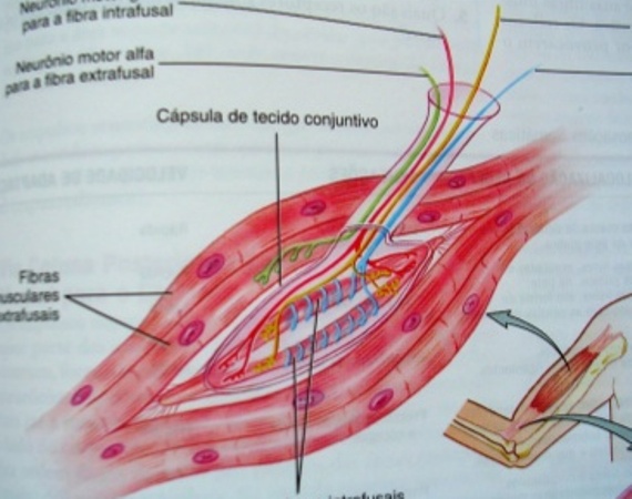
Are modified muscle fibers contained in a capsule of connective tissue that attach to and have parallel extrafusal fibers. The central portion is surrounded by a sensory neuron, being unable to contract. Not so the ends are provided with actin and myosin proteins and are innervated by neurons thinner than conventional engines muscle fibers, these are called motor neurons range. (MONTEIRO and FARINATTI, 2000).
Each muscle spindle is formed by about 4 to 20 small specialized muscle fibers, called intrafusal fibers (within the zone), which is present encased in a connective tissue sheath. In turn, the intrafusal fibers are controlled by specialized motor neurons, called motor neurons range and extrafusal fibers (regular muscle fibers away from the spindle) are innervated by alpha motoneurons (WILMORE e COSTILL, 2001 apud GREGO NETO, 2007).
The spindle basically has the function of informing the change in velocity and the extension (length) muscle. Under these conditions respond to any number of elongation (GUYTON; HALL, 1997).
When the muscle is elongated, the central portion follows the movement by activating the sensory neuron to send impulses to the spinal cord where synapses with the alpha motor neurons that once stimulated, sends commands towards the stretched muscle fibers to contract and shorten muscle stimuli and decreasing the flow coming from zone (MONTEIRO; FARINATTI, 2000).
Image reference: slideshare
 Are modified muscle fibers contained in a capsule of connective tissue that attach to and have parallel extrafusal fibers. The central portion is surrounded by a sensory neuron, being unable to contract. Not so the ends are provided with actin and myosin proteins and are innervated by neurons thinner than conventional engines muscle fibers, these are called motor neurons range. (MONTEIRO and FARINATTI, 2000).
Each muscle spindle is formed by about 4 to 20 small specialized muscle fibers, called intrafusal fibers (within the zone), which is present encased in a connective tissue sheath. In turn, the intrafusal fibers are controlled by specialized motor neurons, called motor neurons range and extrafusal fibers (regular muscle fibers away from the spindle) are innervated by alpha motoneurons (WILMORE e COSTILL, 2001 apud GREGO NETO, 2007).
The spindle basically has the function of informing the change in velocity and the extension (length) muscle. Under these conditions respond to any number of elongation (GUYTON; HALL, 1997).
When the muscle is elongated, the central portion follows the movement by activating the sensory neuron to send impulses to the spinal cord where synapses with the alpha motor neurons that once stimulated, sends commands towards the stretched muscle fibers to contract and shorten muscle stimuli and decreasing the flow coming from zone (MONTEIRO; FARINATTI, 2000).
Image reference: slideshare
Are modified muscle fibers contained in a capsule of connective tissue that attach to and have parallel extrafusal fibers. The central portion is surrounded by a sensory neuron, being unable to contract. Not so the ends are provided with actin and myosin proteins and are innervated by neurons thinner than conventional engines muscle fibers, these are called motor neurons range. (MONTEIRO and FARINATTI, 2000).
Each muscle spindle is formed by about 4 to 20 small specialized muscle fibers, called intrafusal fibers (within the zone), which is present encased in a connective tissue sheath. In turn, the intrafusal fibers are controlled by specialized motor neurons, called motor neurons range and extrafusal fibers (regular muscle fibers away from the spindle) are innervated by alpha motoneurons (WILMORE e COSTILL, 2001 apud GREGO NETO, 2007).
The spindle basically has the function of informing the change in velocity and the extension (length) muscle. Under these conditions respond to any number of elongation (GUYTON; HALL, 1997).
When the muscle is elongated, the central portion follows the movement by activating the sensory neuron to send impulses to the spinal cord where synapses with the alpha motor neurons that once stimulated, sends commands towards the stretched muscle fibers to contract and shorten muscle stimuli and decreasing the flow coming from zone (MONTEIRO; FARINATTI, 2000).
Image reference: slideshare